Research & Publications > The Hong Kong Practitioner > Clinical Quiz

Clinical Quiz (Please login 'Member Area' for online submission of latest issue)
Clinical Quiz December 2016
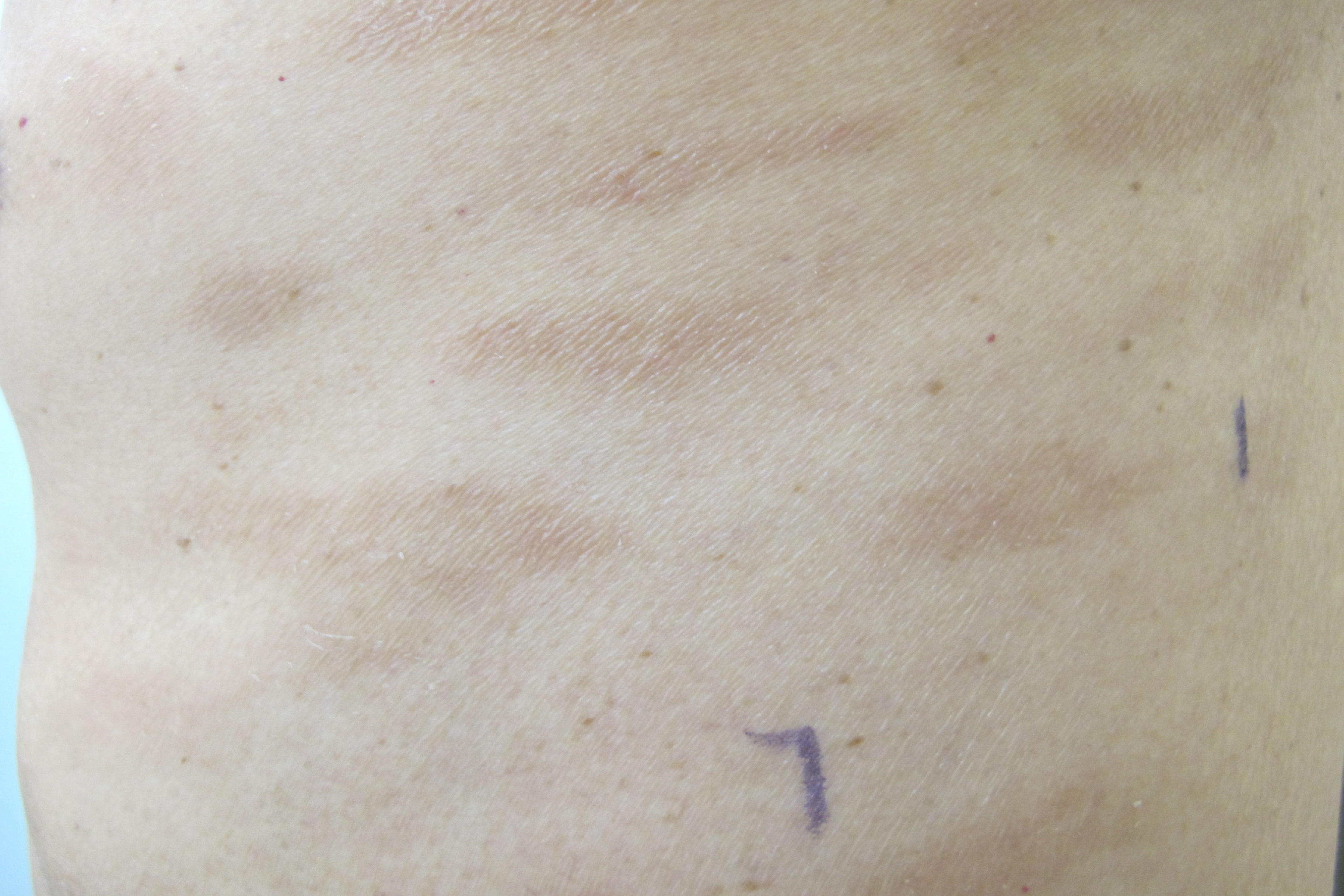
This gentleman presented with long standing non itchy rashes on his body
King-man Ho
|
Readers are invited to participate in the Clinical Quiz. A prize draw, sponsored by Pfizer Corporation Hong Kong Limited, will be undertaken among the successful entries. For entry into the draw, simply answer the question, fill in the reply slip and return it to the College or submit the answer through the website by 23 February 2017. Each reader is allowed to submit one entry only. The name of the winner and the answer will be published in the March 2017 issue.
|
Clinical history:
This gentleman presented with long standing non itchy rashes on his body that was not responsive to topical 0.025% fluocinolone acetonide cream.
What is the clinical diagnosis?
| A. | Chronic superficial scaly dermatitis |
| B. | Tinea incognito |
| C. | Mycosis fungoides |
| D. | Pityriasis versicolor |
Answer:
A. Chronic superficial scaly dermatitis
Clinical examination of this gentleman revealed multiple brownish fairly well demarcated elongated patches with very fine wrinkling on the surface located on the trunk.
Chronic superficial scaly dermatitis (CSSD) is also named as chronic superficial dermatitis, Brocq disease, small plaque parapsoriasis, or digitate dermatitis. CSSD starts as asymptomatic macules with fine superficial scaling on the trunk or flexural regions of limbs. Lesions may gradually increase in size and number. The rashes may slowly become elongated with axis aligned to the skin lines and thus the condition has also been named as “digitate” dermatitis. The borders are usually not very clearly defined. Yellowish hue has described in textbook written by authors in Caucasian countries, and the term xanthoerythodermia perstan is coined to name this condition. A slightly tanned hue is however more commonly observed in Chinese. The wrinkling on the surface may lead to an impression of skin atrophy, but histologically as there is no epidermal thinning, this is described as pseudoatrophy. The coarse pattern may sometimes lead to an impression of lichenification, however there is no actual thickening of skin. It is described as pseudolichenification by some authors. The nature and cause of CSSD is not yet clear. At some stage, the term “parapsoriasis” was coined with a view to group CSSD with “large plague” parapsoriasis that is now thought to be a precursor or even early lesion of mycosis fungoides (MF). Nowadays, the prevailing belief is that CSSD is a benign disease with, if any, minimal risk of progression to MF and thus may be an entity distinguished from “large plague parapsoriasis”. CSSD has a chronic disease course that may sometimes be better in the summer months perhaps because of sunlight exposure though the trunk is usually covered.
Tinea incognito refers to dermatophyte infection of the skin but without the typical features of tinea. Tinea incognito happens when dermatophyte infection involves those immunosuppressed hosts. It is not uncommonly encountered after using topical steroid treatment for tinea misdiagnosed as eczema or other dermatological diseases. As the body inflammatory response is attenuated, tinea incognito may not be symptomatic. The clues to alert the clinician of the diagnosis of tinea incognito include the history of long term use of topical or systemic steroid, presence of skin atrophy in and around the lesions, persistence or even progression of disease lesions despite initial improvement of itchiness and an otherwise clear cut tinea infection in vicinity of the lesion. Skin scraping for fungal study is required to confirm the diagnosis.
Mycosis fungoides (MF) is a specific type of T cell lymphoma of the skin. The typical lesions of patch stage mycosis fungoides are patches with variable and bizarre shapes with predilection to trunk, buttock and proximal limbs. Both intralesional and interlesional variation in morphology that means for example strict border in one quadrant and circular or concave border at the other borders, and roundish lesion on one site but other bizarre shaped lesions on the other sites may be noticeable. Scale may be present and typically fine. Poikilodermatous (atrophy, telangiectasia and dyspigmentation) changes may be present. Itchiness is variable. Skin biopsy with or without T cell receptor gene rearrangement is required to confirm the diagnosis.
Pityriasis versicolor is a superficial infection by pityrosporum yeast, Malassezia furfur. It presents with multiple fairly well demarcated hypopigmented macules with predilection to chest and upper back sometimes extended also to neck, upper arm and face. It may also present with tanned instead of hypopigmented macules especially in those people with light colour complexion. Fine yellowish superficial scale may be present in some lesions that confer the descriptive term “pityriasis” for this skin infection. Pityriasis versicolor are more commonly encountered during the summer months that may be explained by excessive body oil secretion in the hot weather that facilitate growth of the yeast or tanning of the uninfected skin that exaggerate the contrast between skin lesions with infection and uninfected surrounding skin. Diagnosis is mainly clinical. Wood’s light examination may assist the differentiation from lesions with depigmentation such as in vitiligo. Skin scraping with wet mount microscopy may confirm the diagnosis.
The clues to suspect CSSD in this patient are long standing asymptomatic digitate lesions on the trunk. Skin scraping for wet mount microscopic examination and skin biopsy were performed to exclude pityriasis versicolor and MF in this patient.
Apart from skin care and simple emollients to relieve dryness and scaling, treatment is not required in asymptomatic patients. Topical steroids may be used to reduce itching if it happens in the involved patient. Controlled sun exposure may be advised or even phototherapy may be tried for those who are particular concerned of the lesions. Phototherapy may be effective to clear the lesions temporarily.
The slide and the question were prepared by:
Dr King-man Ho, FRCP (Glasg, Edin), MRCP (UK), FHKCP, FHKAM (Medicine)
Consultant Dermatologist-in-Charge,
Social Hygiene Service, PHSB, CHP, DH
Back
