Research & Publications > The Hong Kong Practitioner > Clinical Quiz

Clinical Quiz (Please login 'Member Area' for online submission of latest issue)
Clinical Quiz September 2023
A 39-year-old woman has known history of systemic lupus erythematosus (SLE) on hydroxychloroquine (HCQ) and prednisolone
Sze-man Wong
|
Readers are invited to participate in the Clinical Quiz*. Simply answer the question, fill in the reply slip and return it to the College by 17 November 2023. Each reader is allowed to submit one entry only. The name of the winner and the answer will be published in the December 2023 issue.
*Note: There would be no prize award for this issue while sponsorship for Clinical Quiz has been ended in September 2020 issue. The answer of the Clinical Quiz for this issue will be announced in the next issue. Thank you for your support.
|
Clinical history:
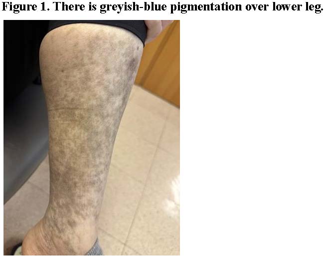
A 39- year-old woman has known history of systemic lupus erythematosus (SLE) on hydroxychloroquine (HCQ) and prednisolone. She presented with blue-greyish discoloration over lower legs (Figure 1) for 3 months. She denied of other over-the-counter medications or herbal medicine.
What is cause of the discoloration of lower legs?
| A. | Melasma |
| B. | Post inflammatory hyperpigmentation |
| C. | HCQ-related hyperpigmentation |
| D. | Ashy dermatosis |
Answer:
C. HCQ-related hyperpigmentation
Melasma is a commonly acquired skin disorder that presents as bilateral, blotchy, brownish pigmentations most commonly distributed over the face. Extrafacial involvement includes the forearms, upper arms, shoulder sun-exposured areas, and rarely over the lower legs. Melasma is more common in women than in men, with an onset typically between the ages of 20 and 40 years. The cause of melasma is complex; it has been proposed that it is a photoaging disorder in genetically predisposed individuals. The pigmentation generally results from the overproduction of melanin by melanocytes; either taken up by keratinocytes in the epidermal melanosis or deposited in the dermis forming melanophages in dermal melanosis. About 60% of cases report affected family members. Sun exposure, and hormonal surge such as the use of oestrogen/ progesterone containing oral contraceptive pills and pregnancy has been implicated in the development of melasma. The use of Wood’s lamp and dermoscopy can be used to differentiate epidermal and dermal melasma. Clinically, epidermal melasma are likely more well defined with brownish pigmentation and more obvious on Wood’s lamp examination; dermal melasma are likely less defined, more blue-grey discoloration and show no accentuation upon Wood’s lamp examination.
Post-inflammatory hyperpigmentations are nonspecific skin changes which can occur after resolution of any inflammatory condition in the skin especially in people of a darker skin colour.
Ashy dermatosis, also known as, erythema dyschromicum perstans is characterised by well circumscribed round to oval or irregular patches on the face, neck and trunk that are grey in colour. It is a form of acquired dermal macular hyperpigmentation. The pigmented patches can be symmetrical in distribution or unilateral. Early lesions may be reddish in colour, often with a more pronounced border and elevated. It is not always observed and patients are otherwise well with no associated disease or blood test abnormality.
Ashy dermatosis may occur at any age but it appears to be more prevalent in young adults. Women are affected more than men. It is more resistant to treatment and persists for years although some may disappear on its own.
In this specific patient, given the background of SLE and HCQ intake, distribution over bilateral lower legs, with pigmentation gradually fading upon the discontinuation of HCQ, the mostly likely diagnosis is of HCQ-related hyperpigmentation. The incidence of HCQ-related hyperpigmentation in patients with SLE is estimated to be about 5%. Potential risk factors include bruising, corticosteroid use, oral anticoagulants, antiplatelet agents, skin trauma and sun exposure.1 A skin biopsy from the lesion can show granular pigment deposition with positive Fontana– Masson staining for melanin and Perls’ Prussian blue staining for iron, which confirms the diagnosis of HCQ induced hyperpigmentation.2
The hyperpigmentation usually gradually fades after discontinuation of the drug, but complete clearance is rare. Reassurance of the benign nature of this pigmented condition and the use of a trial of topical steroid and Q-switched ruby laser could result in the regression of these lesions.
References:
1. Jallouli M, et al. Plaquenil Lupus Systemic Study Group. Hydroxychloroquine-induced pigmentation in patients with systemic lupus erythematosus: a case-control study. JAMA Dermatol. 2013 Aug;149(8):935-940.
2. Muller R, Jarrot PA, Kaplanski G. Hydroxychloroquinerelated hyperpigmentation. Rheumatology (Oxford). 2022 Feb 2;61(2):873.
The slide and the question were prepared by:
Dr. Sze-man Wong, MBBS, MRCP(UK), MSc(London), FHKCP, FHKAM, FRCP(Edin)
Consultant,
Division of Dermatology, Department of Medicine, Queen Mary Hospital, Hong Kong SAR
Back
