Research & Publications > The Hong Kong Practitioner > Clinical Quiz

Clinical Quiz (Please login 'Member Area' for online submission of latest issue)
Clinical Quiz March 2023
An 81-year-old man presented with right groin rash for 1 year
Ka-chun Cheng, Sze-man Wong
|
Readers are invited to participate in the Clinical Quiz*. Simply answer the question, fill in the reply slip and return it to the College by 17 May 2023. Each reader is allowed to submit one entry only. The name of the winner and the answer will be published in the June 2023 issue.
*Note: There would be no prize award for this issue while sponsorship for Clinical Quiz has been ended in September 2020 issue. The answer of the Clinical Quiz for this issue will be announced in the next issue. Thank you for your support.
|
Clinical history:
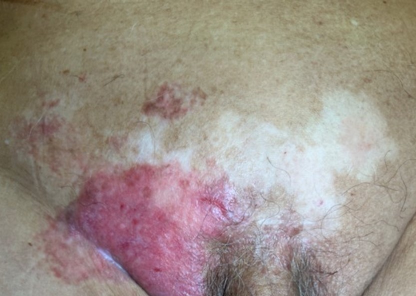
An 81-year-old man presented with right groin rash for 1 year. The rash was otherwise asymptomatic, not pruritic nor painful. He had a history of symptomatic abdominal aortic aneurysm with emergency endovascular aneurysm repair (EVAR) 8 months ago. He received coronary artery stenting after an episode of non-ST elevation myocardial infarction (NSTEMI) 7 months ago. The right groin lesion was noted by the vascular surgeons during his routine vascular clinic follow up. Clinically there was a well-demarcated erythematous plaque over the right groin region, with some background of vitiligo-like hypopigmented patches at the pubic region. There was no other skin/nail/scalp involvement. His groin lesion progressively expanded despite treatment with topical 1% clotrimazole cream and topical corticosteroid . He denied venereal exposure over the past 2 years.
What is the diagnosis?
| A. | Atypical mycobacterial infection |
| B. | Inverse psoriasis |
| C. | Cutaneous candidiasis |
| D. | Extramammary Paget disease |
| E. | Squamous cell carcinoma |
Answer:
D. Extramammary Paget disease
The clinical photo showed a well-demarcated erythematous plaque with white scales over the right
groin. There were a few smaller erythematous papules and plaques located superior to largest plaque. A depigmented patch at the pubic area, which was present for more than 20 years, represents a background of vitiligo. There was no palpable groin lymph node. There was no lesion over his penis, scrotum, perineum, and anal area.
A 4mm-punch skin biopsy performed over his right groin confirmed the diagnosis of extramammary
Paget disease (EMPD). Immunostaining of Cytokeratin 20 (CK20) negative and Gross cystic disease fluid
protein-15 (GCFPD-15) positive favours cutaneous origin.
His prostate-specific antigen (PSA) was not elevated. His screening colonoscopy done 6 months ago was unremarkable. Cystoscopy showed no bladder tumour.
Wide local excision was performed with a curative intent.
EMPD is a rare intraepithelial carcinoma arising from sites rich in apocrine glands such as the groins, vulva, scrotum, penis, and perianal area. Clinicians should be vigilant and suspected a diagnosis of EMPD for patients presenting with a well-demarcated erythematous plaque with whitish scales over the groins or anogenital area. Erosions and white scale on an erythematous plaque give rise to the classic “strawberries and cream” appearance. EMPD may become invasive and metastasize via the lymphatic system if not treated early.
EMPD can be primary or secondary. Immunostaining with positive CK20 is highly associated with internal malignancies. Secondary EMPD may arise from an underlying visceral malignancy such colorectal or urogenital malignancies. A thorough cutaneous and lymph node examination should be performed. Pelvic and breast examination should be performed in women and prostate examination in men. Further investigations including colonoscopy, cystoscopy, mammogram in women and prostate-specific antigen in men is required, especially for lesions demonstrating positive CK20. A local study demonstrated 8.3% of EMPD cases were associated with an internal malignancy.1
Treatment is mainly by wide local excision with or without reconstruction or Mohs micrographic surgery if expertise is available. A local study showed the recurrence rate was 14.6%.1 Other treatment options including radiotherapy, topical imiquimod, topical 5-fluorouracil, photodynamic therapy, and CO2 laser ablation. Patients should have long-term follow-up to monitor for any recurrence.
For this case, the lesion was refractory to topical antifungal and topical corticosteroid. Clinicians should reconsider alternative diagnoses. Important diagnoses such as cutaneous malignancy or atypical skin infection must be ruled out. Investigation such as skin scrapping for fungal smear and culture, and skin biopsy will be essential in this case.
The slide and the question were prepared by:
Dr. Ka-chun Cheng, MBBS(HK), MRCP(UK), MSc GEOR(CUHK), FHKCP, FHKAM(Medicine)
Specialist in Geriatric Medicine
Associate Consultant,
Department of Medicine, Queen Mary Hospital, Hong Kong SAR
Dr. Sze-man Wong, MBBS(HK), MRCP(UK), MSc(London), FHKCP, FHKAM(Medicine), FRCP(Edin)
Specialist in Dermatology and Venereology
Consultant,
Division of Dermatology, Department of Medicine, Queen Mary Hospital, Hong Kong SAR
Back