Research & Publications > The Hong Kong Practitioner > Clinical Quiz

Clinical Quiz (Please login 'Member Area' for online submission of latest issue)
Clinical Quiz June 2017
This elderly woman presented with asymptomatic lesions on her lower lip for a few years
King-man Ho
|
Readers are invited to participate in the Clinical Quiz. A prize draw, sponsored by Pfizer Corporation Hong Kong Limited, will be undertaken among the successful entries. For entry into the draw, simply answer the question, fill in the reply slip and return it to the College or submit the answer through the website by 24 August 2017. Each reader is allowed to submit one entry only. The name of the winner and the answer will be published in the September 2017 issue.
|
Clinical history:
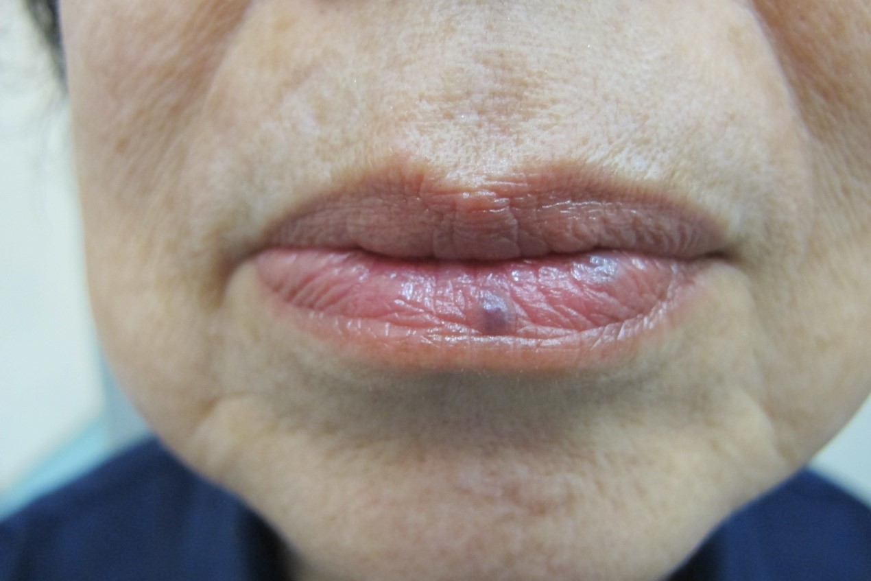
This elderly woman presented with asymptomatic lesions on her lower lip for a few years. The lesions slowly increased in size initially and remained stable.
What is the clinical diagnosis?
| A. | Venous lake |
| B. | Oral naevus |
| C. | Labial melanotic macule |
| D. | Melanoma |
Answer:
A. Venous lake
The clinical photo showed a bluish red slightly elevated lesion on the mid portion of lower lip. Another similar lesion was present on the left side of lower lip.
Venous lake presents with an asymptomatic small soft compressible dark blue slightly elevated lesion in an elderly person. The blood content in venous lake can be emptied upon persistent pressure showing its “venous nature”. It involves the lower lip more than the upper lip. The superficial skin crease on the lip is not disturbed. Occasionally if the blood inside the lesion is thrombosed, it becomes firm and remains dark blue in colour arousing the suspicion of a melanocytic lesion. Venous lake sometimes also presented in other part of the face including ear.
Oral naevus results from benign proliferation of naevus cells in the epithelial, submucosal (dermis in skin), or both layers like its counterpart of acquired melanocytic naevus of skin. It presents with asymptomatic light brown and dome shaped elevated lesion on the lip, and other part of the oral mucosae. Comparing to the prevalence of acquired melanocytic naevi of skin, it seems that oral naevus is very uncommon. Despite the well described oral mucosal melanoma, the risk of malignant change of oral naevus has not been well described and probably low. Blue naevus with its deeper pigmented cellular component can also occur in oral mucosa, it may appear dark bluish in colour.
Mucosal melanotic macule can be regarded as lentigo simplex (“lentigines” is the plural of “lentigo”) involving the mucosae including oral and genital regions. Histopathologically, lentigo is characterized by an increase in melanocytes but without forming nest in the basal layer of the oral mucosal epithelium. Therefore in contrast to acquired melanocytic naevus, it is flat clinically. Labial melanotic macule presents with asymptomatic flat brownish to darker coloured macules on the lip and with surface skin creases undisturbed. Lesions are sometimes not very clearly demarcated and usually measured few mm in size. Peutz-Jeghers syndrome is one of the well-known syndromes that may present with multiple lentigines on the lip. Carney complex presents with multiple lentigines on the face sometimes also the lip atrial myxomas, and selected endocrine hyperactivity.
Melanoma of the oral mucosa arises from malignant change of melanocyte of the basal layer of the mucosa. Primary oral melanoma is not common except perhaps in Japan. No definite risk factors are so far identified. Excessive sun exposure and sunburn as risk factors for melanoma on the skin is not applicable in oral mucosa. Oral melanoma remains asymptomatic till progression has occurred. Some may report a change in a pre- existing pigment lesion. Up to one third of oral melanoma are pertained to amelanotic melanoma that presents with white, mucosa-coloured or red mass. The palate and maxillary gingiva are involved in about 80% of primary melanoma whereas mandible, tongue, and buccal mucosa are the site of predilection for metastatic melanoma. Though blue naevus is more common on the palates, malignant change of blue naevus has not been reported. As general principles in recognition of malignant lesion, a lesion with colour variegation, ulceration, satellite lesions and association of regional lymph node enlargement should arouse one to the possibility of malignancy. The principles are applicable for oral melanoma.
The colour, consistency, blanchable nature, and undisturbed surface creases and location on the lower lip help to differentiate venous lake to other pigment lesions. Further investigation is not required.
As venous lake is a benign and asymptomatic lesion, proactive treatment is not required. If treatment is contemplated, simple excision or other destructive modalities such as electrocauterisation are the options.
The slide and the question were prepared by:
Dr King-man Ho, FRCP (Glasg, Edin), MRCP (UK), FHKCP, FHKAM (Medicine)
Consultant Dermatologist-in-Charge,
Social Hygiene Service, PHSB, CHP, DH
Back
