|
May 2003, Volume 25, No. 5
|
Case Report
|
||
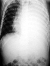
A 5-year old Chinese boy with pulmonary tuberculosis that presented with pleural effusionK K Chan 陳強杰, D K K Ng 吳國強, K L Kwok 郭嘉莉, P Y Chow 周博裕, W F Lau 劉永暉 HK Pract 2003;25:231-233 Summary Pulmonary tuberculosis (PTB) is endemic in Hong Kong. The presenting symptoms can be non-specific. We present a 5-year old boy who had been coughing for 6 weeks in association with a low-grade fever. He was diagnosed to have PTB only after chest radiograph showed a pleural effusion. Early diagnosis would have been possible if a chest radiograph and Mantoux test had been done earlier. 摘要 肺結核病是香港的一種地方性流行病。它的症狀可以是非特異性的。我們報告一個五歲的男孩,咳嗽六個星期並有低熱,最後發現胸部x光顯示朐腔積液,而診斷為肺結核。如果及早照肺和做 Mantoux 試驗就可以早做出診斷。 Introduction In 1999, there were 7512 cases of tuberculosis (TB) notified in Hong Kong, 81 (1%) cases were reported in children aged less than 15 years old.1 Classical systemic symptoms include fever, night sweats, anorexia, weight loss and malaise. Organ-specific symptoms include persistent cough, pleuritic pain, and haemoptysis.2 The diagnosis of TB infection in children depends primarily on a high index of suspicion and appropriate investigations. It is suggested that TB infection should be considered in any child who has had a persistent cough and fever for more than two weeks unresponsive to antibiotic treatment.3 Around the world, almost 500 children die of tuberculosis (TB) infection every day. It has been estimated that 170,000 children die each year from the two most severe forms of disseminated disease, meningeal and miliary TB.4 In order to control the spread of TB, a high index of suspicion, accurate diagnosis, adherence to full course of treatment and contact tracing are all necessary. We report a child with pulmonary tuberculosis (PTB) that presented with a persistent cough for six weeks. The pulmonary tuberculosis was complicated by pleural effusion. The diagnosis of PTB was established by pleural biopsy. Case report
WSF was a five-year old boy. He was born in Mainland China and came to Hong Kong
three years ago. His parents claimed that his immunisation schedule was completed
in China and Hong Kong though no Bacille Calmette-Guerin (BCG) scar was noticed.
He enjoyed good past health and lived with his parents and elder brother in a 100-square-feet
rented room in Shamshuipo. He presented with loss of appetite, malaise, night sweats,
cough and low grade fever (oral temperature: 38
Upon admission, he was lethargic. His temperature was 38.4
Anti-TB drugs including isoniazid, rifampicin and pyrazinamide were prescribed for 6 months. Prednisolone was given for a month because of the presence of pleural effusion. The fever settled three days later. Chest drain was removed two weeks later with a total of 975ml of pleural fluid collected. Repeat CXR showed only minimal pleural effusion. There was no derangement of liver enzymes after the use of anti-TB drugs. The case was notified to the Department of Health and the whole family was referred to the Chest Clinic for family screening. Chest x-rays of his parents were normal. His elder brother had a positive Mantoux test with 20 x 21mm induration but there was no evidence of TB disease. Discussion This case illustrates the non-specificity of symptoms of TB infection and the importance of appropriate investigations for the diagnosis. The diagnosis of TB infection in children depends primarily on a high index of suspicion. Clinicians should be alert to the possibility of PTB when faced with a child who complains of persistent cough. In this case, the diagnosis would have been made earlier if proper physical examination, CXR, complete blood count including ESR and Mantoux Test had been performed earlier. Pleural effusion is not uncommon in TB infection. By three months of pulmonary TB infection, 25% of cases developed a pleural effusion and by six months, there would be an effusion in 75% of cases.5 Effusions are usually unilateral but can be bilateral.6 Pleural effusion results from a hypersensitivity reaction to TB proteins and liposaccharides rather than microbial invasion of the pleura. Less than 10% of cases have positive AFB stain of pleural fluid as the bacilli seed from a subpleural pulmonary focus or a casesated lymph node. Culture of pleural fluid grew Mycobacterium tuberculosis in fewer than 65% of cases. In contrast, histological examination and culture of pleural tissue increase the diagnostic yield to between 80% and 90%.7,8 Thus chest tapping coupled with pleural biopsy increase the yield of TB identification in pleural effusion. It was clearly demonstrated in this case as M.tuberculosis was only recovered in the pleural biopsy. Conclusions Medical practitioners should be aware of tuberculosis infection as a differential diagnosis when they encounter a child with persistent cough for more than two weeks. Pleural biopsy should be attempted before insertion of chest drain when tuberculosis infection is suspected. Key messages
K K Chan, MBBS
Medical Officer, D K K Ng, MMedSc, FRCP, FHKAM(Paed) Consultant Paediatrician, K L Kwok, MBChB, MRCP, FHKAM(Paed) Senior Medical Officer, P Y Chow, MBChB, MRCP, DCH Medical Officer, W F Lau, MBChB, MRCP, FHKCPaed Specialist Medical Officer, Department of Paediatrics, Kwong Wah Hospital. Correspondence to : Dr D K K Ng, Department of Paediatrics, Kwong Wah Hospital, Kowloon, Hong Kong.
References
|
|||